


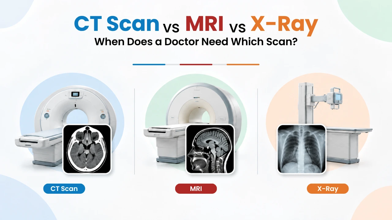
X-ray, CT and MRI in one minute
All three scans let a doctor look inside your body without surgery. They differ in what they use (radiation or a magnet), what they see best, and how long they take.
X-ray
Bones · chest · first check
A tiny burst of radiation passes through you and makes one flat (2D) picture. Bones show up white, so breaks are easy to spot.
- Uses :- Radiation (low dose)
- Time :- A few seconds to minutes
- Best for :- Fractures, chest, joints, arthritis
CT scan
Emergencies · organs · complex bone
You lie on a table that slides through a ring. An X-ray beam spins around you and a computer builds thin slices, like slicing a loaf of bread.
- Uses :- Radiation (higher dose)
- Time :- About 5–10 minutes
- Best for :- Head injury, stroke, bleeding, chest, abdomen
MRI
Brain · spine · nerves · ligaments
You lie inside a tunnel-shaped magnet. Strong magnetism and radio waves create very detailed pictures of soft tissue. It is noisy but painless.
- Uses :- Magnet + radio waves (no radiation)
- Time :- About 20–60 minutes
- Best for :- Slipped disc, nerves, brain, ligaments, cartilage
CT scan vs MRI vs X-ray: key differences
Here is the difference between an X-ray, a CT scan and an MRI scan side by side.
Swipe left to see all columns
| Feature | X-ray | CT scan | MRI scan |
|---|---|---|---|
| How it works | One beam of X-rays, one flat picture | Many X-rays from all angles, built into 3D slices | Strong magnet + radio waves |
| Radiation | Very low | Moderate (more than an X-ray) | None |
| Time on the table | Seconds | About 5–10 minutes | About 20–60 minutes |
| Sees best | Bones, lungs | Bones in detail, organs, bleeding, blood vessels | Brain, spinal cord, discs, nerves, muscles, ligaments |
| Emergency use | Fractures | First choice — very fast | Rarely first — too slow |
| Noise / comfort | Quiet, quick | Quiet, open ring | Loud; narrow tunnel; must lie still |
| Metal implants | No problem | No problem | Must be checked first |
| Relative cost | Lowest | Medium | Highest |
Which scan for which problem?
This is the question most people really want answered. The table below shows the scan a doctor usually reaches for first, and when they may move to another one. Your doctor may choose differently after examining you — that is normal.
Swipe left to see all columns
| Problem or symptom | Usual first scan | May be followed by | Why |
|---|---|---|---|
| Head injury after a fall or accident | CT | MRI later, if symptoms continue | CT finds bleeding and skull fractures in minutes |
| Suspected stroke (face drooping, weak arm, slurred speech) | CT | MRI | CT quickly rules out a brain bleed before treatment starts |
| Suspected broken bone | X-ray | CT for complex or joint fractures | Most breaks show clearly on an X-ray |
| Low back pain (no red flags) | Often no scan at first | MRI if pain lasts 6+ weeks or leg weakness appears | Most back pain settles without a scan |
| Pain shooting down the leg (sciatica), slipped disc | MRI | — | Only MRI clearly shows discs pressing on nerves |
| Knee giving way, locking, sports injury | X-ray | MRI | X-ray checks bone; MRI shows ligament, meniscus and cartilage |
| Joint pain in an older adult (arthritis) | X-ray | Rarely needs more | X-ray shows joint space narrowing well |
| Seizures, long-term headaches, memory problems | MRI | — | MRI shows brain tissue in far more detail |
| Cough, fever, breathlessness | Chest X-ray | CT chest | X-ray spots pneumonia; CT shows the lungs in detail |
| Severe tummy pain, kidney stone | Ultrasound or CT | CT | CT is very good at finding stones and bowel problems |
| Lump, or checking a known cancer | CT or MRI | Depends on the organ | Liver, brain and pelvis often need MRI; chest and abdomen often CT |
Why doctors often start with an X-ray (and not an MRI)
Many patients ask, "Why did my doctor give me an X-ray? Shouldn't I get an MRI?" It is a fair question. Doctors usually work step by step, for three good reasons:
The simplest test often gives the answer
If you hurt your wrist, an X-ray shows a break in seconds. An MRI would show more detail, but it would not change the plaster or the treatment.
Less radiation, less cost, less waiting
An X-ray is cheap, quick and widely available. Starting there saves you money and time when it is enough.
An MRI can show things that are not the problem
Many adults have disc bulges or "wear and tear" on an MRI that cause no pain at all. Scanning too early can lead to worry — or even treatment — for something harmless. The scan should match your symptoms.
CT scan vs MRI: how does a doctor choose?
CT scan vs MRI is the comparison people search for most. The short answer: CT is chosen for speed and bone detail; MRI is chosen for soft-tissue detail.
A doctor usually picks a CT scan when…
- It is an emergency — head injury, accident, suspected stroke or internal bleeding.
- They need fine detail of bones, such as a complex fracture or the spine after a fall.
- They need to see the lungs, chest or abdomen — for example pneumonia, kidney stones or appendicitis.
- You cannot have an MRI — because of a pacemaker, some metal implants, or severe fear of closed spaces.
- You cannot lie still for long because of pain or illness.
A doctor usually picks an MRI when…
- They need to see the brain or spinal cord in detail — seizures, long-term headaches, MS, tumours.
- You have back or neck pain with leg or arm symptoms, like a slipped disc pressing on a nerve.
- They suspect a torn ligament, meniscus or cartilage in the knee or shoulder.
- They want to avoid radiation — for example in children, young adults needing repeat scans, or (with care) in pregnancy.
MRI scan vs CT scan: when you need both
Sometimes you need both. A CT may be done first in an emergency, and an MRI later to look more closely. That is not a wasted scan — each one answers a different question. This is the main difference between an MRI scan and a CT scan in practice.
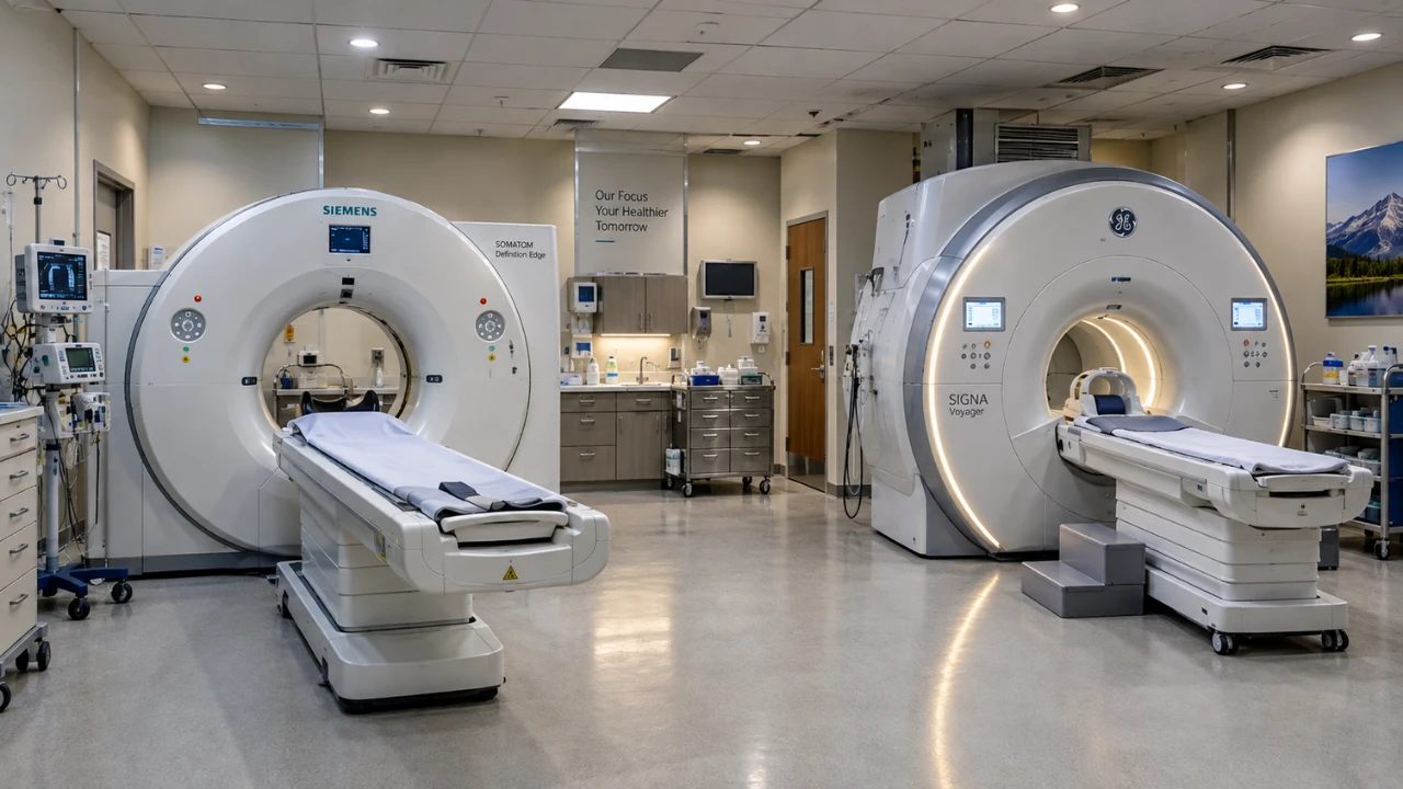
CT scan vs X-ray: what's the difference?
A CT scan is a kind of X-ray — but a much more powerful one. The difference between an X-ray and a CT scan is like a single photo versus a full 3D model.
- An X-ray squashes everything into one flat picture. Organs overlap, so small problems can hide behind bones.
- A CT scan takes hundreds of pictures from all around you. The computer separates them into slices, so nothing hides.
That extra detail comes with more radiation and a higher cost. So in the CT vs X-ray choice, doctors use an X-ray when a simple picture is enough, and a CT scan when they need to see what the X-ray cannot — such as a hairline fracture in the spine, bleeding inside the head, or a small lung spot.
How much radiation is in each scan?
We all get small amounts of natural radiation every day from soil, air and even food. Scan doses are often compared to this "background" radiation. These are typical adult figures from the American College of Radiology and RSNA [1]:
Swipe left to see all columns
| Scan | Typical dose | Same as natural radiation over about… |
|---|---|---|
| Chest X-ray | 0.1 mSv | 10 days |
| Spine X-ray (lower back) | 1.4 mSv | 6 months |
| CT head | 1.6 mSv | 7 months |
| CT chest | 6.1 mSv | 2 years |
| MRI (any part) | 0 | No radiation |
So is a CT scan dangerous? For one scan that you need, the benefit is almost always far bigger than the very small risk. The risk adds up with many scans over a lifetime, which is why doctors avoid CT scans that are not needed — especially for children [2]. Keep a record of your past scans and show it to your doctor.
Who should not have an MRI or a CT scan?
Check before an MRI if you have:
- A pacemaker or defibrillator — some newer ones are "MRI-safe", but many are not. The team must check the device card.
- Metal in the body — such as a cochlear implant, aneurysm clip, metal fragments in the eye (common in welders and metal workers), or a nerve stimulator. Most joint replacements, plates and screws are safe, but always tell the team .
- Fear of closed spaces (claustrophobia) — tell the team in advance. They can explain the steps, let a relative stay nearby, or arrange mild sedation.
- Pregnancy — MRI is generally preferred over CT in pregnancy when a scan is truly needed, but contrast dye is usually avoided. Always say if you might be pregnant.
Check before a CT scan if you:
- Are or may be pregnant — CT of the belly is avoided unless it is an emergency.
- Need contrast dye (an injection that makes blood vessels and organs show up clearly) and have kidney disease, diabetes, or a past allergy to dye. You may need a kidney blood test (creatinine) first.
How to prepare for your scan
- Bring your doctor's prescription and old reportsOld X-ray, CT or MRI films and CDs help the radiologist compare and spot changes.
- Tell the team about metal, devices and pregnancyPacemaker, implants, clips, metal fragments — and whether you may be pregnant.
- Ask if you need to fastSome CT and MRI scans with contrast need 4–6 hours without food. Confirm the hospital's own instructions.
- Bring a recent kidney test if contrast is plannedEspecially if you are over 60, diabetic or have kidney problems.
- Wear loose clothes with no metalRemove jewellery, hairpins, watches and belts. You may be given a gown.
- Bring a family member if you are anxiousIt helps with claustrophobia and with getting home if you were sedated.
What changes the cost of a scan?
In India, an X-ray is usually the cheapest scan, a CT costs more, and an MRI costs the most. The exact price depends on:
- The type of scan — X-ray, CT or MRI.
- The body part — one knee costs less than the whole spine.
- Plain or contrast — a scan with contrast dye costs more than a plain scan.
- Special studies — such as CT angiography (blood vessels) or detailed MRI protocols.
- Emergency or planned — and whether insurance or a TPA covers your scan.
Please call the Aditya Hospital radiology desk for the current price of your scan.
Scans at Aditya Hospital, Rewari
You do not need to travel to Gurgaon or Delhi for routine or emergency imaging. Aditya Hospital's radiology department in Rewari offers:
- Digital X-rayClear images within seconds.
- Multi-slice CT scanFast scanning of the head, chest, abdomen, bones and blood vessels.
- MRIBrain, spine and joint imaging. Confirm in-house MRI and field strength.
- Ultrasound & DopplerRadiation-free imaging for the abdomen, pregnancy and blood vessels.
- 24×7 emergencyCT support for head injury, stroke and trauma. Confirm CT is available round the clock.
- Same-day reportsMost reports are ready the same day, read by a radiologist.
Reports are checked by our radiologist, Dr. Saranya Manikandan. If your scan shows a brain or spine problem, you can see our neurosurgery & spine team in the same hospital. For bone and joint injuries, our orthopaedics department can review your X-ray or MRI.
Frequently Asked Questions
Q1. Which is better, a CT scan or an MRI?
Neither is better overall. A CT scan is better for emergencies, bones, the lungs and bleeding because it is fast. An MRI is better for the brain, spinal cord, nerves, discs and ligaments because it shows soft tissue in more detail without radiation. The better scan is the one that answers your doctor's question.
Q2.MRI scan vs CT scan: which is safer?
An MRI uses no radiation, so it avoids that risk. But it is not safe for everyone — people with some pacemakers or metal implants cannot have one. A CT scan uses a small amount of radiation; for a needed scan, the benefit is much bigger than the risk.
Q3.Why would a doctor order a CT scan instead of an MRI?
Usually because it is an emergency and speed matters, because they need fine bone detail, because they are looking at the lungs or abdomen, or because you cannot have an MRI due to a device, metal implant or claustrophobia.
Q4. Is a CT scan just a stronger X-ray?
In a way, yes. Both use X-rays. But an X-ray takes one flat picture, while a CT scan takes many pictures from all around you and builds them into 3D slices. That shows far more detail, with a higher radiation dose.
Q5.CT vs X-ray: which is used for a fracture?
An X-ray is almost always first. Most broken bones show clearly on it. A CT scan is added for complex fractures, fractures near a joint, the spine or the face, or when the X-ray looks normal but a break is still suspected.
Q6. Do I need an MRI for back pain?
Usually not at first. Most back pain improves within a few weeks. An MRI is advised if the pain lasts more than about 6 weeks despite treatment, or straight away if you have leg weakness, numbness, or loss of bladder or bowel control — the last is an emergency.
Q7. Can I have an MRI if I have a knee replacement or metal plate?
Most modern joint replacements, plates and screws are safe for MRI. But you must tell the team, and bring any implant card. Pacemakers, cochlear implants, some brain clips and metal pieces in the eye need special checks.
Q8. How long does a CT scan and an MRI take?
A CT scan usually takes about 5–10 minutes on the table. An MRI usually takes about 20–60 minutes, depending on the body part and whether contrast is used. An X-ray takes only seconds.
Q9. Is an MRI scan or a CT scan more expensive in India?
An MRI scan is usually more expensive than a CT scan, and both cost more than an X-ray. The price depends on the body part and whether contrast dye is used. Call the radiology desk at Aditya Hospital for the current price.

Dr. S.N. Manikandan
Dr. Manikandan leads the Neurosurgery & Spine department at Aditya Hospital and uses CT and MRI scans of the brain and spine to plan treatment for his patients. Client to confirm exact qualifications and years of experience before publishing.


